|
Back to Annual Symposium Program
Ultrasound Navigation for Endovascular Aortic Intervention
Gabriel Herscu, M.D., Jay Mung, M.S., John Moos, M.D., Sukgu Han, M.D., Grace Huang, M.D., Jesse Yen, Ph.D., Fred Weaver, M.D..
University of Southern California, Los Angeles, CA, USA.
OBJECTIVES:
Radiation exposure and nephrotoxic contrast injections are inherent to conventional endovascular treatment regimens in aortic disease. Contrast nephropathy is estimated to occur in up to 50% of patients undergoing administration of contrast. Radiation exposure during endovascular aortic intervention is significant and the long-term effects have not been fully realized. Our objective was to determine navigational precision of this novel technique.
METHODS:
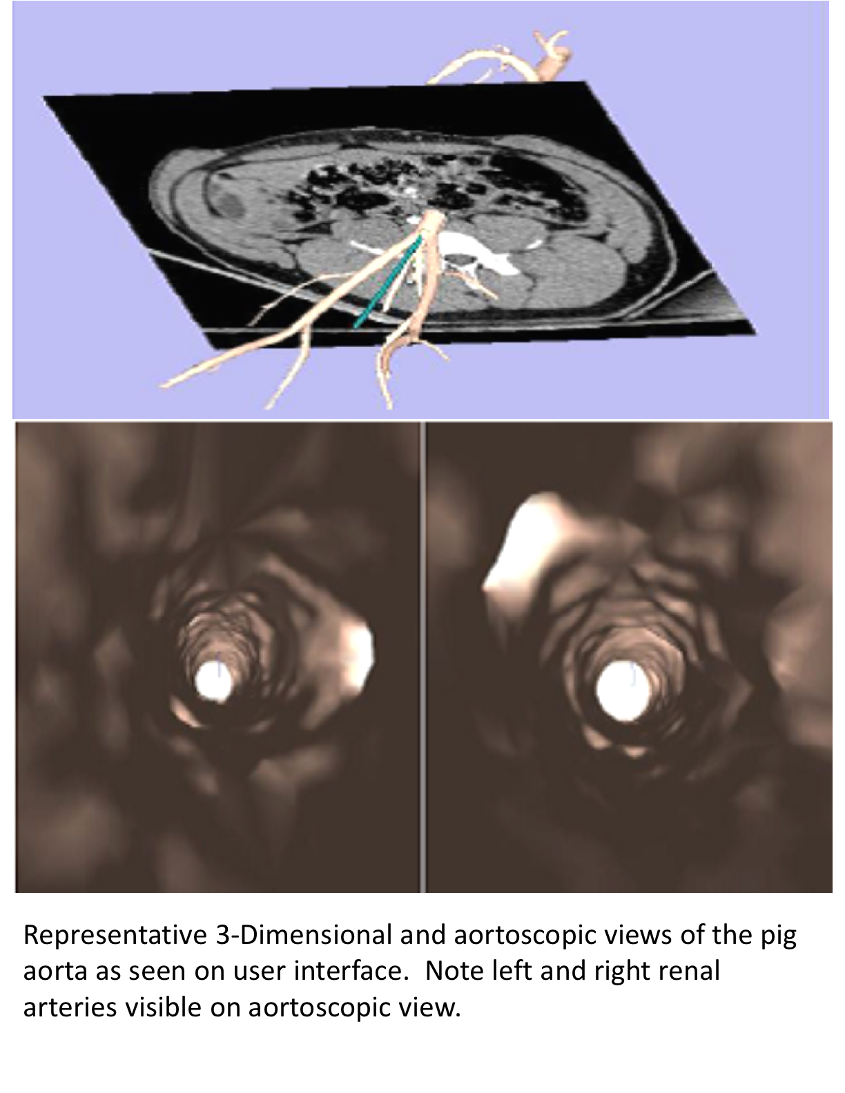
We designed a navigational system for placement of aortic endovascular prostheses using an ultrasound guidance system with a graphical user interface (GUI). Our system utilizes an endovascular ultrasound transmitter passed into the aorta on the tip of a catheter and continuously tracked via trilateration with external ultrasound receivers. Graphical representation of catheter location coupled in 3-D with preoperative CTA is represented on a monitor along with a virtual aortoscopic view looking forward from the catheter tip. This procedure was performed in a pig model. Movement of the catheter via ultrasound guidance was compared using correlation plot with fluoroscopic measurements. Ultrasound-guided catheter movement was also compared to aortic centerline as determined by preoperative CT A. After data acquisition, a covered, self-expanding stent was advanced and deployed at the inferior edge of the right renal artery as determined by the ultrasound guidance system. The pig was then sacrificed and the aorta opened in-situ to evaluate accuracy of stent placement in relation to the right renal artery orifice.
RESULTS:
Compared with fluoroscopy data, ultrasound navigation showed excellent concordance (RMS = 0.6mm, R2 > 0.99 ). Tracking of catheter position showed a mean difference of 2.15mm when compared to aortic centerline for all recorded catheter tip positions. At aortic dissection, the stent was found within 2mm of the renal orifice.
CONCLUSIONS:
Ultrasound navigation in endovascular aortic intervention is feasible and precise when compared to fluoroscopic catheter manipulation. Its virtual-reality graphical user interface allows intuitive, real-time manipulation of endovascular devices, while avoiding the damaging effects of radiation and contrast administration.

Back to Annual Symposium Program

|