|
Back to Annual Symposium Program
A Light-based, Radiation-free Angiographic Simulator for the Study of Complex Hemodynamics
Doran Mix, B.S.1, Daniel B. Phillips, Ph.D.2, Steven Day, Ph.D.2, Nicole Varble, M.S.1, Karl Q. Schwartz, M.D.1, Ankur Chandra, M.D.1.
1University of Rochester, Rochester, NY, USA, 2Rochester Institute of Technology, Rochester, NY, USA.
OBJECTIVE: The experimental study of hemodynamics through angiography is difficult. Most studies require large animal models with concurrent exposure to ionizing radiation in dedicated, expensive animal facilities. Our goal was to develop an experimental angiographic system without the need for ionizing radiation and animal models.
METHODS: A physiologically accurate, in vitro arteriovenous fluid model was used to simulate the vascular system. An Infimed high-resolution CCD imaging unit was used to image the photo-lucent vascular model over a light source (Figure 1A). Black photo-opaque dye was used as the contrast medium in varying concentrations delivered through a power injector. The entire imaging system was driven through a customized computer interface.
RESULTS: The use of the light-based system allowed for both fluoroscopic (Figure 1B) and digital-subtraction DICOM image (Figure 1C) acquisition and storage to a local PACS server for future study. Total imaging area obtained was 506cm2 with spatial 1024x1024 pixel resolution of 250μm2 per pixel at a maximum frame rate of 15 fps. Various parameters including flow, contrast density, and downstream contrast dilution were quantified after varying contrast injection rates. The system accurately imaged real-time catheter and guidewire movements during physiologic flows and contrast injections.
CONCLUSIONS: Through the novel utilization of imaging technology and in vitro hemodynamic simulation, experimental angiographic studies can be carried out free of radiation and large animal work in a variety of anatomic situations. The current limitations include the inability to image through tissues, requiring recreation of the vascular system of interest on the hemodynamic simulator. The potential applications of this technology include training, device testing, and development of angiographically-derived hemodynamic algorithms which would be too cumbersome or unethical to obtain in human or animal settings. 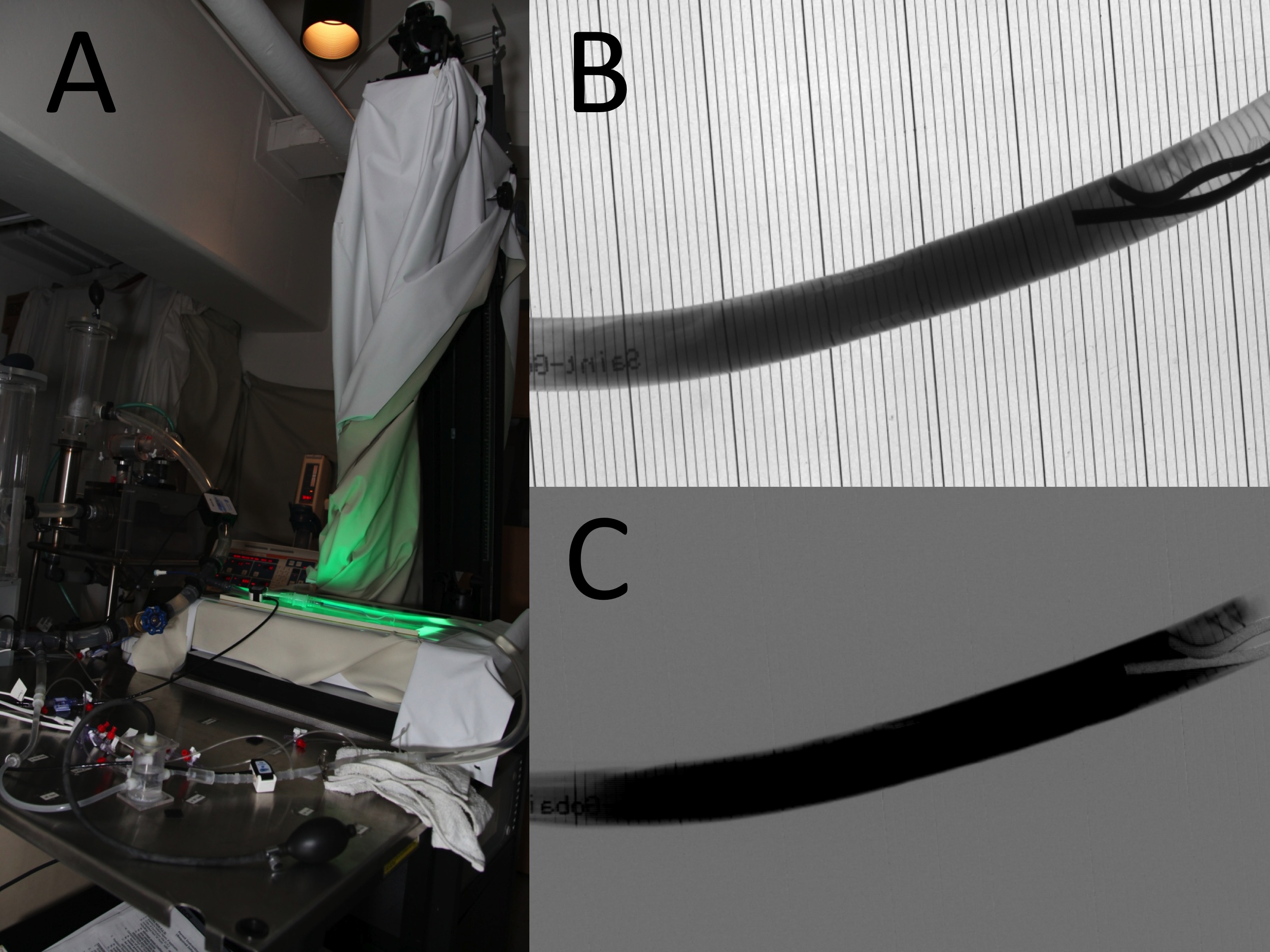
Back to Annual Symposium Program

|

