Back to Annual Symposium Program
Centerline Three Dimensional Reconstruction versus Axial Computed Tomography Angiography for Sizing of Endovascular Grafts in Patients with Abdominal Aortic Aneurysm
Marlene T. O'Brien, MD, PhD1, Adam J. Doyle, MD1, Jennifer L. Ellis, MD1, Karl A. Illig, MD2, Ankur Chandra, MD1.
1University of Rochester, Rochester, NY, USA, 2University of South Florida, Tampa, FL, USA.
OBJECTIVE: Correct sizing of endovascular aortic grafts is essential for successful elective treatment of infrarenal abdominal aortic aneurysms (AAA) and for decreasing the rate of delayed secondary interventions. Our study compares the reproducibility and accuracy of sizing aortic endografts using centerline 3D reconstruction as compared with standard computed tomography angiography (CTA) axial images.
METHODS: Patients who underwent elective endovascular infrarenal aneurysm repair (EVAR) from 2006-2010 who had axial and centerline images were selected for retrospective review. Power analysis to be able to detect a 2 mm difference between measurement methods was conducted and revealed a total of 122 studies would need to be reviewed. Six diameter and three length measurements were compared using two-tailed t-test and Pearson correlation coefficients. The agreement between the two methods was analyzed using Bland and Altman’s method. EVAR main body graft selection based on proximal neck measurements obtained by both sizing methods was also compared using a Chi-square test.
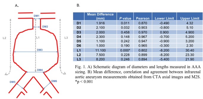
RESULTS: Axial and centerline images and measurements from 124 patients were reviewed. The mean difference between diameters was not statistically significant (p>0.001), with acceptable agreement at all points except at L1 (11.1 mm, p=0.00). Pearson correlation coefficients were above 0.90 at all diameter measurements apart from DM1 (0.870). EVAR main body graft sizing based on the largest neck diameter was studied for axial versus centerline, with no statistical difference between the graft selected using either method (p=0.980).
CONCLUSIONS: AAA measurements obtained from CTA axial images are similar to those obtained from centerline 3D reconstruction images based on six aortic diameter measurements and three lengths. EVAR main body grafts selected based on axial or centerline images demonstrated no statistical difference between the two methods. Use of CTA axial images alone may be adequate for selection of main body graft selection in patients with infrarenal AAA.
Back to Annual Symposium Program

|
