|
|
 |
Back to Annual Symposium Program
Aortic hemodynamics post thoracic endovascular aortic repair: bird-beak drawback
Guido H. W. van Bogerijen1, MD, Ferdinando Auricchio2, 3, PhD, Michele Conti2, PhD, Adrien Lefieux3, Alessandro Reali2, PhD, Alessandro Veneziani4, PhD, Tiziano Passerini4, PhD, Santi Trimarchi1, MD, PhD
1IRCCS Policlinico San Donato, Thoracic Aortic Research Center, University of Milan, Milan, Italy, 2Department of Civil Engineering and Architecture (DICAr), Pavia University, Pavia, Italy, 3CESNA Center for Advanced Numerical Simulations, Institute for Advanced Study of Pavia (IUSS), Pavia, Italy, 4Department of Mathematics and Computer Science, Emory University, Atlanta, GA
Objective
Thoracic endovascular aortic repair (TEVAR) frequently involves a proximal landing zone in the inner curvature of the distal aortic arch, potentially causing a bird-beak configuration. Bird-beak has been associated with endoleaks after TEVAR. This study quantitatively evaluates impact of TEVAR on aortic hemodynamics, with special focus on bird-beak.
Methods
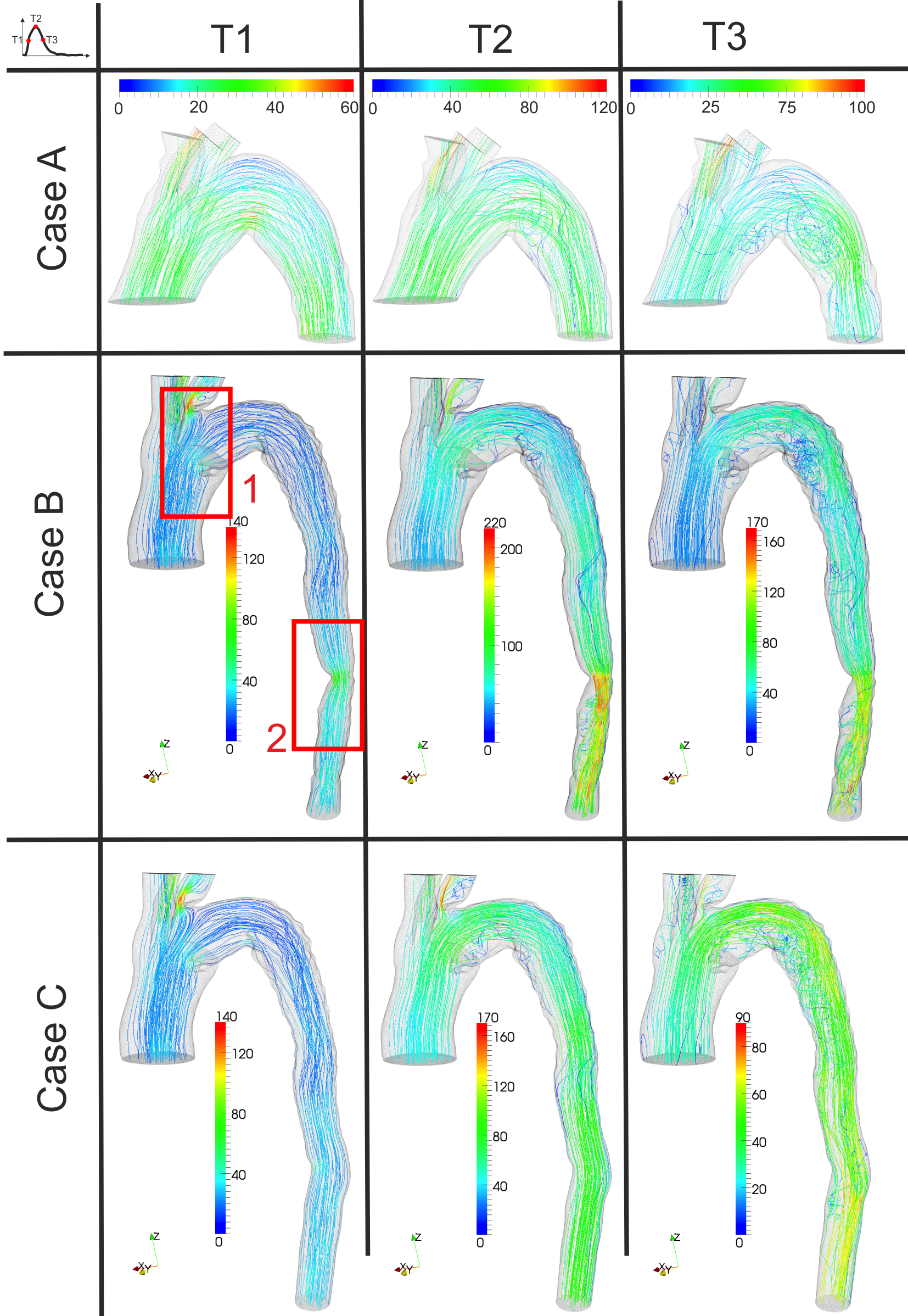
Pre- and postoperative computed tomography angiography from a patient treated with TEVAR for post-dissecting thoracic aneurysm was used to evaluate the anatomical changes induced by TEVAR and to generate the computational network essential for computational fluid dynamics (CFD) analysis. This analysis was focused on bird-beak configuration, flow distribution in supra aortic branches with (partial) coverage of the origin of the left subclavian artery (LSA) and stenosis in the distal descending thoracic aorta due to compression of the true lumen by the false lumen. Three different CFD analyses, cases A-C, (A=preoperative lumen; B=postoperative lumen; C=postoperative lumen, computed without stenosis) were performed and compared with each other at three time points T1-T3 (T1=maximum acceleration of blood flow; T2=systolic peak; T3=maximum deceleration of blood flow).
Results
Postoperatively, disturbance of flow is reduced at the bird-beak location due to change of geometry after TEVAR when comparing A and B. The stent graft protrusion with partial coverage of the origin of the LSA produces disturbance of flow in the LSA. Strong velocity increase and flow disturbance at the location of aortic stenosis compared to no stenosis are found when comparing B and C; however stenosis in the distal descending aorta has no effect on the aortic arch hemodynamics. (Figures 1,2)
Conclusions
CFD can help physicians in understanding change in aortic hemodynamics after TEVAR, giving useful information about effects of bird-beak, coverage of the LSA and a narrowed true lumen.
Figure 1. Velocity streamlines for investigated cases (A-C) at three time points (T1-T3). The corresponding velocity (cm/s) is used to color each streamline.
Figure 2. Contour plot of pressure (mmHg) distribution along a cross-section in the bird-beak region for case B.

Back to Annual Symposium Program

|
