Back to 2016 Annual Symposium ePosters
Superficial Femoral Artery Stent Fracture Resulting in Pseudoaneurysm: A Case Report and Review of Literature
Maham Rahimi, MD, PhD, Lynn Doctor, MD, Jonathan Bath, MD.
University of Cincinnati, Cincinnati, OH, USA.
INTRODUCTION
Arterial pseudoaneurysms have different etiologies, including trauma, iatrogenic injury, following vascular reconstruction, infection, and vasculitides. In this case, a mechanical fall caused fracture of a superficial femoral artery (SFA) stent, leading to mid-thigh pseudoaneurysm.
METHODS
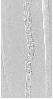
A 73 year old male patient presented with left thigh pain after falling from his wheelchair. His past medical history is unreconstructable coronary artery disease, COPD, left SFA stenting in 2009 and left ventricular thrombus on Warfarin. He was found to have a painful large left mid-thigh pulsatile mass. Left thigh radiography revealed SFA stent fracture (Figure 1). Duplex ultrasound demonstrated a 5.0 x 5.6 cm pseudoaneurysm with active flow. Further workup revealed MRSA bacteremia and intravenous antibiotics were instituted.
RESULTS
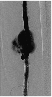
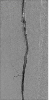
Due to high cardiopulmonary risk with an open bypass procedure, an endovascular approach was favored. Diagnostic angiography revealed large pseudoaneurysm (Figure 2). Initially 6 mm x 40 mm EverFlex stent was placed to allow preferential flow into the SFA stent leading to thrombosis of the pseudoaneurysm sac. Repeat duplex demonstrated pseudoaneurysm persistence and a 6 x 100 mm rifampin soaked Viabahn stent was then placed. Completion angiography revealed elimination of pseudoaneurysm sac (Figure 3). Routine follow-up duplex demonstrated sac thrombosis without signs of infection.
CONCLUSION
Stent fracture is of unclear consequence and in coronary arteries has been rarely complicated by pseudoaneurysm formation. We reported a very unusual case of SFA stent fracture with large symptomatic pseuodaneurysm in a bacteremic patient. Although open bypass and interval ligation is favored in the setting of bacteremia the patient was at very high risk for open surgery. This case study highlights a rare complication of SFA stenting and to our knowledge is the only report of SFA stent fracture causing symptomatic pseudoaneurysm in the literature.
Figure 1. Left thigh X-ray revealed SFA stent fracture

Figure 2. Diagnostic angiogram revealed flow through the fractured region with the pseudoaneurysm sac

Figure 3. Completion angiography revealed continuous flow throughout the SFA without any filling of the pseudoaneurysm sac

Back to 2016 Annual Symposium ePosters
|


