|
Back to 2016 Annual Symposium ePosters
Validating Subclavian Artery Duplex Ultrasound Peak Systolic Velocity with Angiographic Defined Stenosis
Albeir Y. Mousa, MD1, Mike Broce, BA2, Androw Sticco, MD1, Ravi Viradia, MD1, Shane Monnett, MD1, Michael Yacoub, MD1, Mark Bates, MD1, Ali AbuRahma, MD1.
1R C Byrd Health Sciences Center, W. Va. Univ., Charleston, WV, USA, 2CAMC Research Institute, Charleston, WV, USA.
OBJECTIVES:
To date, no consensus exists for duplex ultrasound (DUS) criteria in the diagnosis of significant subclavian artery stenosis. In our current practice, peak systolic velocity (PSV) values of 250 cm/sec are used to identify 70% stenosis, but these values have not been validated. The purpose of our study was to investigate the correlation between PSV values and stenosis, while the main objective was to determine the cut-off point for predicting significant angiographic defined subclavian stenosis (>70%).
METHODS:
A retrospective chart review was conducted based on a convenience sample of patients evaluated for suspected subclavian or carotid stenosis with available subclavian DUS and angiograms from 2000 to 2014. Pearson’s correlation and a receiver operating characteristics curve (ROC) analysis were used to compare PSV and calculated angiographic measurement of subclavian stenosis based on the North American Symptomatic Carotid Endarterectomy Trial (NASCET) criteria.
RESULTS:
In total, 214 arteries in 123 patients were evaluated, of which 53% were female with an average age of 65 +12 years. Twenty-three arteries had retro-grade flow, it was found that 20 out of the 23 retro-grade flow arteries had significant angiographic stenosis (>70%), while the remaining three had stenosis (>50%). The remaining 191 arteries had ante-grade flow and were deemed suitable for accurate PSV measurements. There was a moderate significant correlation between PSV and calculated stenosis (R=0.63, p<0.001). PSV >240 cm/sec was found to be the optimal cut-off point for predicting angiographic defined subclavian stenosis (>70%) with an area under the curve (AUC = 0.85, 95%CI: 0.77-0.93, p<0.001). In addition, the PSV >240 cm/sec cut-off point produced a sensitivity of 75.8% and specificity of 82.3%, with corresponding 47.2 and 94.2% positive and negative predictive values, respectively. Overall accuracy was 81.2%.
CONCLUSIONS:
Establishing subclavian duplex criteria to characterize significant stenosis is crucial to identify patients that require further imaging modalities or treatment. Retro-grade vertebral flow was an absolute indicator of underlying subclavian occlusive disease. In our accredited laboratory, subclavian PSV >240 cm/sec was found to be a threshold that can be used to identify significant angiographic defined (>70%) subclavian stenosis with a high degree of accuracy. 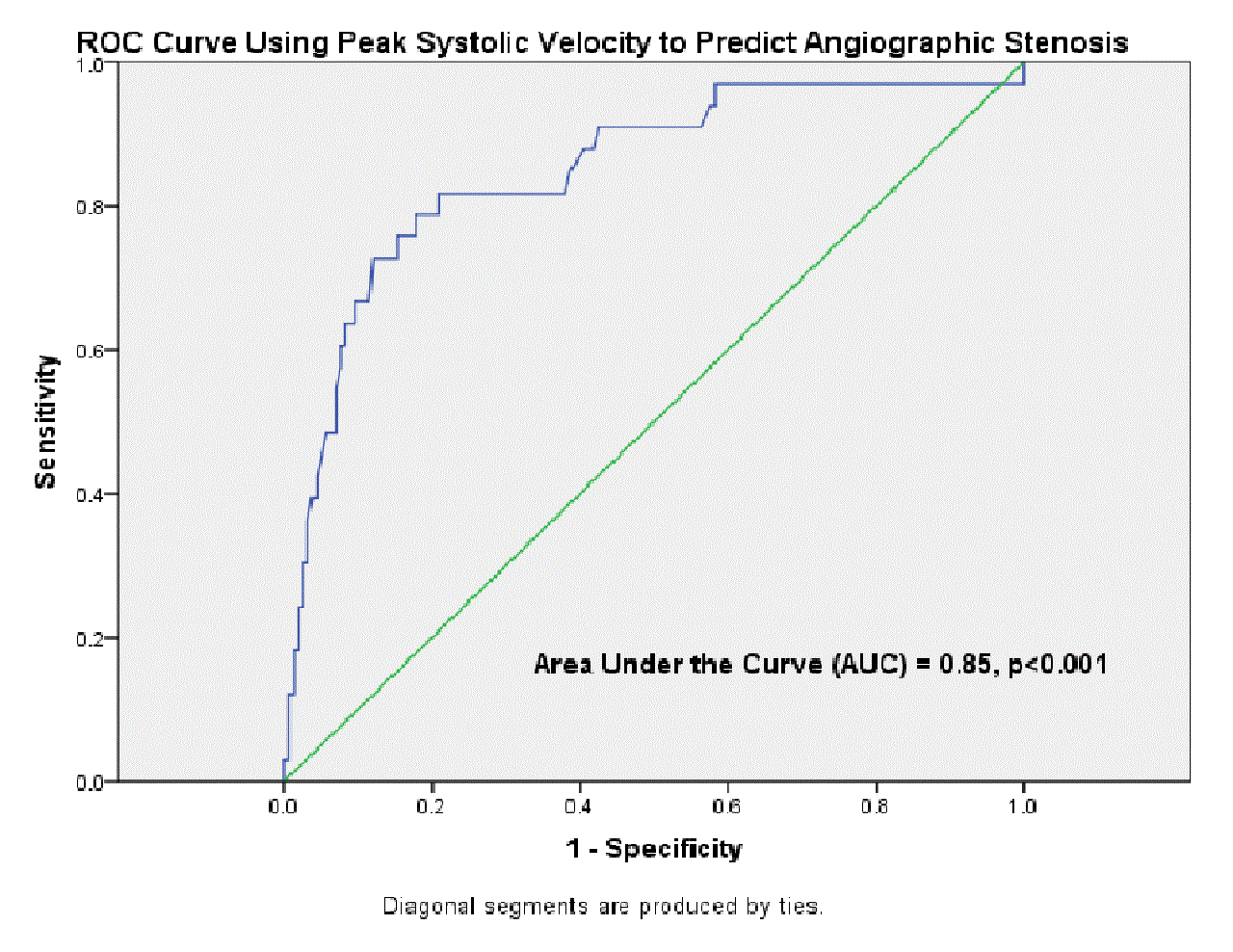
Back to 2016 Annual Symposium ePosters
|


