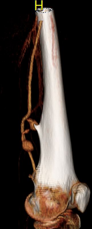
Vascular complications from hereditary multiple osteochondromas: A case report and review of literature
Sonia Talathi, M.D., Reshma Duffy, Larry Scher.
Montefiore Medical Center, New York, NY, USA.
Objective: Osteochondromas are the most common benign bone tumors, occurring either alone or as part of the autosomal dominant syndrome hereditary multiple osteochondromas. Osteochondromas are usually asymptomatic, with a 4% complication rate. Vascular complications are rare and must be taken urgently to the operating room. We present a case report and literature review of these complications.
Methods: A new case of superficial femoral artery (SFA) pseudoaneurysm from osteochondroma is described followed by a review of literature. A bibliographic search was performed online (PubMed, ScienceDirect) from 1955 to 2016 using the descriptions "osteochondroma and vascular complications, pseduoaneurysms and exostoses/osteochondroma."
Results: Our patient is an otherwise healthy 59-year-old female who presented with two days of left medial thigh pain and swelling, with no history of trauma. She had a family history of hereditary multiple osteochondromas (HMO). The patient underwent a CT angiogram, which revealed a left SFA pseudoaneurysm measuring approximately 3x2 cm with a large femoral osteochondroma adjacent to the arterial injury. The patient was taken to the operating room for open surgical repair and excision of the osteochondroma through a medial thigh incision. The femoral osteochondroma had partially avulsed a geniculate branch of the lateral SFA with active bleeding seen. The injury was closed primarily and the osteochondroma was removed. The patient did well with no postoperative complications.
Conclusion: Complications of osteochondromas were first reported in 1955. Only 57 cases involving the lower extremity have been reported and only 10% of all osteochondromas are associated with HMO. Although the most common reported site of injury is the popliteal artery, our patient had an SFA pseudoaneurysm. Our patient was also significantly older than the typical presentation seen in young patients approaching skeletal maturity. Controversy exists as to the pathophysiology of vascular complications. Although some authors attribute local trauma to the loss of cartilage, leading to bone spike and eventual vessel perforation, others have demonstrated radiographic changes of osteochondromas prior to symptoms, therefore attributing the loss of cartilage to pressure necrosis from the enlarging pseudoanuerysm. Although rare, vascular complications from osteochondromas may require urgent vascular intervention and resection of the osteochondroma.
Back to 2017 ePosters


