
Interactive Virtual Reality Assisted Evaluation of Inferior Vena Cava Filters For High Risk Retrieval
Philip L. Auyang, MD1, Vishwanath Chegireddy, MD1, Demet Gurlur, PhD2, Alan B. Lumsden, MD, FACS1.
1Houston Methodist Hospital, Houston, TX, USA, 2EchoPixel, Mountain View, CA, USA.
OBJECTIVES: The purpose of this study is to investigate the utility of interactive virtual reality (IVR) true 3D imaging to assess inferior vena cava (IVC) filters for high risk retrieval. Tertiary centers receive a substantially large volume of referrals for IVC filter removal, often after previous failed attempts at endovascular retrieval. Filter fracture, tilt, erosion/perforation, migration, and thrombosis can cause significant morbidity and potentially interfere with endovascular retrieval. IVR is a novel platform for pre-operative assessment of IVC filters and may provide insight into stent integrity (i.e. fracture) and location to adjacent structures which may otherwise not be well appreciated on conventional CT viewing.
METHODS: We retrospectively reviewed 12 patients who underwent complex IVC filter retrieval from August 2016 to September 2017. Pre-operative CT images were viewed on EchoPixel workstation (EchoPixel, Mountain View, CA) in an IVR environment. Qualitative comparisons were made with operative reports.
RESULTS: IVR correctly identified 4 out of 4 fractured filters upon verification with operative reports. IVC filters were extracted with open surgical excision (7/12) and endovascular retrieval (5/12). True 3D IVR viewing provided the means to accurately assess compromising stent geometry such as fracture, tilt, and protrusion. These defects were immediately noticeable upon IVR viewing while 2D CT required continued panning to develop a mental 3D projection (Figure 1). With immediate 3D reconstruction this also eliminated the need for post processing, thus saving time. Other notable findings were the visibility of location of filter components in adjacent structures. Two filter components were found in the right renal vein and one in the right gonadal vein which was difficult to appreciate on conventional CT viewing.
CONCLUSIONS: With the increasing volume of IVC filter placement, growing complications from longer than intended indwelling time, and ongoing litigations, the volume of complex IVC filter retrieval will surely increase over the coming decade. Pre-operative assessment with IVR can aid surgical planning by confidently detecting filter component fractures. Identifying involvement with adjacent structures can assist with surgical planning and exposure. Additionally, identifying filter component erosion may be possible with endoluminal transform function capabilities for which ongoing investigation is taking place. 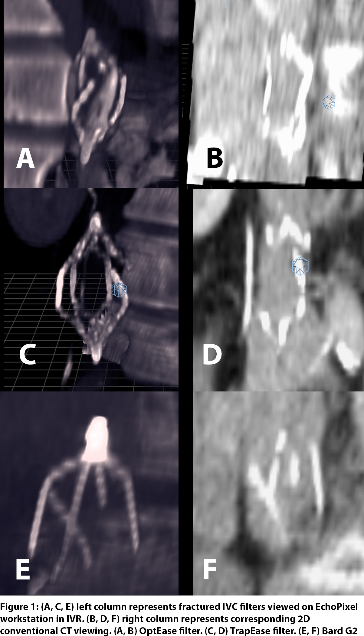
Back to 2018 ePosters


