
Progressive Stenosis of a Popliteal Artery Stent Graft by Laminated Thrombus
Ali Rteil, MD, Rob Harriz, MD, Martina Draxler, MD, Ziad Al Adas, MD, Farah Mohammad, MD, Yasaman Kavoousi, MD, Loay Kabbani, MD.
Henry Ford Health System, Detroit, MI, USA.
Objectives: Endovascular repair of Popliteal Artery Aneurysms (PAAs) using self-expanding covered stents is an accepted alternative to open repair. However long-term patency has been shown to be decreased. There are several methods for stent failure. We report a case of in-stent stenosis with laminated thrombus.
Methods: We introduce the case of a 75-year-old male, who presented with PAA at a tertiary care center. He was treated with a Viabahn stent graft (Gore, Flagstaff, AZ). He presented at one-year follow-up new onset claudication. A duplex showed in-stent narrowing. A computed tomography scan showed a significant stenosis within the stent graft, mainly at the level of the knee joint.
Results: Stent graft failures after endovascular PAA repair are well documented. The mechanism of failure include fracture, migration, intimal hyperplasia at the fixation sites. In this case report we hypothesize that layered thrombus developed on the lining of the covered stent with significant stenosis at the level of the knee joint. This layered thrombus formation eventually lead to symptomatic high-grade stenosis.
Conclusion: We describe a new mechanism of developing in stent stenosis in a patient treated for a PAA with a covered self-expanding stent graft. Shedding light on possible mechanisms of stent graft failure in PAA patients may help with the design of new stent grafts that are specific for PAA.
Figure Legend
(a) Pre-op CTA (left) showing Popliteal artery aneurysm with intraluminal thrombus, intraoperative angiogram (center) shows near occlusion of right popliteal artery with distal embolization and intraoperative Angiogram (right) after Stent graft deployment. (b) Angiogram shows narrowing at two points with the knee straight and angiogram with bent knee showing near occlusion of the artery at the proximal stent and narrowing of the artery at flexion points away from the proximal and distal ends.
(c) Duplex ultrasound showing narrowing within the popliteal artery stent graft with flow velocity changes in the stent at 12 months’ post-operatively.
(d) CTA showing near occlusion of the stent graft with laminated thrombus. Hounsfield Units inside and outside stent graft are close in value indicating similar density. 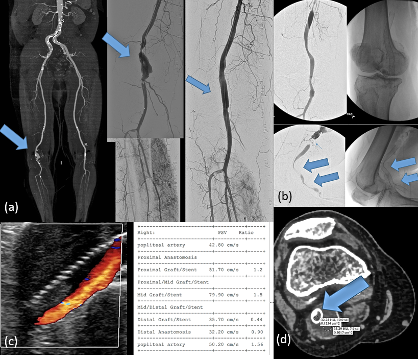
Back to 2019 ePosters