
Endovascular Treatment of Blunt Traumatic Thoracic Aortic Injuries in Aberrant Right Aortic Arch Anatomy
Heepeel Chang, MD, Neal C. Hadro, MD, Marc A. Norris, MD, Alejandro Contreras, MD, Marvin E. Morris, MD.
Baystate Medical Center, University of Massachusetts Medical School, Springfield, MA, USA.
OBJECTIVES: Despite the multiple reported cases of endovascular repair of traumatic thoracic aortic injury, there is paucity of successful endovascular repair of traumatic thoracic aortic injuries in right aortic arch (RAA). We describe a blunt traumatic thoracic injury and successful thoracic endovascular aortic repair (TEVAR) in a patient with a rare type of RAA with bovine variant.
METHODS: Electronic medical records were reviewed to retrieve images, operative details and findings at presentation.
RESULTS: A healthy 34-year old male presented to the emergency department after a blunt trauma to his torso after a motor vehicle accident. He underwent a computed tomography angiography (CTA) that revealed the presence of a RAA with bovine variant as well as a 2x9x9mm pseudoaneurysm located 1 cm distal to the right subclavian artery ostium and mediastinal hemorrhage. He underwent a successful TEVAR with a 24×110mm Valiant® thoracic endograft (Medtronic Inc., Minneapolis, MN) just distal to the right subclavian artery. Final digital subtraction angiography confirmed the complete exclusion of the aortic injury with patent right subclavian artery. A follow-up CTA at 1-year revealed intact repair without evidence of endoleak, kink or fracture of the newly placed stent-graft.
CONCLUSIONS: TEVAR has been widely described in the treatment of thoracic aortic injury with normal aortic arch configuration. Having a blunt traumatic thoracic aortic injury in a patient with aberrant aortic arch anatomy poses unique challenges with paucity of literature describing its use in RAA vessel anomalies. However, our case demonstrates the use of TEVAR as the treatment of choice for blunt traumatic thoracic aortic injury even in RAA with bovine variant.
FIGURE: A. 3D rendering in-situ reconstructed from CTA demonstrating RAA with pseudoaneurysm (arrow) distal to the right subclavian artery. B. Intraoperative digital subtraction arteriography revealing a RAA with a tear distal to the right subclavian artery with a significant pseudoaneurysm (arrow). C. Digital subtraction angiography in a 47° right anterior oblique projection demonstrating successful coverage of the traumatic defect (arrow). 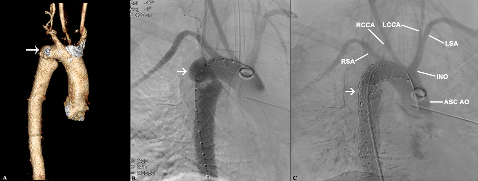
Back to 2019 Abstracts