
Vena Cava Filter With Symptomatic Narrowing and Difficult Reconstruction of IVC and Bilateral Iliac Veins
Whitney Thorne, DO, Brian Kuhn, MD, Matt Recht, MD, Patrick Muck, MD, Aaron Kulwicki, MD.
TriHealth/Good Samaritan Hospital, Cincinnati, OH, USA.
DEMOGRAPHICS:
The patient is a 60 year old Caucasian male current daily smoker with BMI 36.
HISTORY:
Due to anemia the patient was found to have a sigmoid colon mass requiring resection. Prior to resection, the patient developed extensive bilateral lower extremity DVTs. An IVC filter was placed at an outside institution in lieu of anticoagulation due to anemia and upcoming surgery. Four weeks later he was admitted to our hospital with inability to ambulate secondary to worsening edema at which time he was started on anticoagulation. He presented for follow up 6 months later, at which time his DVTs had become chronic. His anticoagulation was stopped, but he was complaining of severe bilateral LE swelling. CTA abdomen demonstrated near occlusion of his infrarenal filter.
PLAN:
Initial trial of conservative measures, including medical grade compression stockings failed after 4 months, due to noncompliance. Then retrieval of the IVC filter from a right internal jugular approach.
DISCUSSION:
The IVC filter was successfully retrieved at which time there was a flow channel through the IVC, therefore we elected to see if his symptoms would improve. He was now compliant with compression but two months later had no improvement. We then proceed with venography via the bilateral CFVs. Wire access into the native vena cava from the right CFV was established, but despite multiple efforts wire access was unable to be established across a chronically occluded left CIV. A Wallstent was then placed extending from the R CIV into the infrarenal IVC with excellent angiographic and IVUS result. A final attempt at crossing the left CIV was made and was successful, however when attempting to pass a catheter into the IVC, the right-sided Wallstent became dislodged and pushed into the suprarenal cava. Using a Coda balloon this was collapsed into an infrarenal position. Then Wallstents were placed in the bilateral CIV and EIV up to the level of the renal veins. Completion angiogram and IVUS demonstrated widely patent stents. The patient was seen at one month follow up with remarkable improvement in his swelling and resolution of his bilateral LE bursting pain. 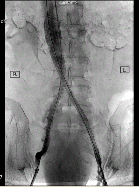
Back to 2019 Abstracts