
Ascending Aortic Access For Imaging And Through-and-through Access Facilitating Endograft Delivery In Complex Aortic Arch Anatomy With Large Subclavian Aneurysm
Celso Uribe, II, MD, William D. Clouse, MD, John A. Kern, MD, Brian P. Fletcher, MD, Stephen Davies, MD.
University of Virginia, Charlottesville, VA, USA.
Introduction: Thoracic aortic arch anatomy and branch aneurysms can pose significant challenges when planning for endograft repair. In this case of a 36 year-old man with large proximal left subclavian aneurysm, our two-step plan of complete arch de-branching followed by femoral delivered TEVAR was antagonized by complex arch angulation. Direct ascending aortic cannulation with through-and-through wire allowed for TEVAR delivery and deployment. 
Case Description: 36 year-old male originally diagnosed with 3cm left subclavian artery aneurysm at age 21. Lost to follow-up. He was transferred to our hospital early May 2019 with complaints of left upper chest pain, syncopal episodes, and a CTA showing an 8.2 cm left subclavian origin aneurysm. He was taken to the OR for aortic arch debranching and TEVAR. Following aortic arch debranching, the patulous subclavian origin and two tortuous arch angles made ascending aortic catheterization from the groin difficult due to wire/catheter bowing into left subclavian artery aneurysm. As a result, we inserted a 7Fr sheath and 5Fr sheath antegrade directly into the ascending aorta via separate purse string cannulations proximal to the debranch graft. Through the 5Fr ascending sheath, arch aortography was possible. Using an EN Snare positioned in the abdominal aorta from the ascending aorta 7Fr sheath, we secured a glidewire from the right femoral sheath and pulled the through ascending aortic sheath. Over this wire, a through-through sheath was placed and the glide wire exchanged for a Lunderquist wire. This resulted in excellent stiff wire position for placement of our two arch endoprostheses. The distal subclavian aneurysm was ligated just proximal to the vertebral artery.
Conclusion: TEVAR delivery and deployment in proximal arch segments may require alternative access. Although direct transaortic delivery from the abdominal aorta is described in cases of peripheral stenosis and small arterial size, it is also possible to use ascending aortic access for imaging and to achieve a stable through-through platform when the arch is tortuous or aneurysm morphology does not allow such from below. 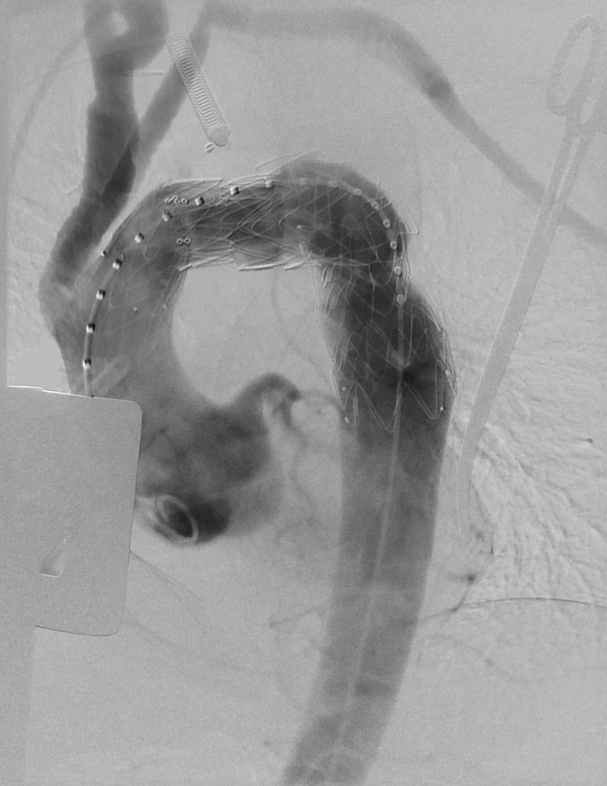
Back to 2020 Abstracts