
Volumetric Measurement Of Abdominal Aortic Calcification And Its Clinical Significance In Dialysis Patients
Itsuo Yokoyama, M.D.1, Tsuyosi Sarai, B.S.R.T.2, Toshinori Asai, M.T.1, Michi Sekikawa, R.N.1, Dage Liu, M.D.1.
1Nagoya Memorial Foundation, Narumi Clinic, Nagoya, Japan, 2Nagoya Memorial Foundation, Shinseikai Daiichi Hospital, Nagoya, Japan.
OBJECTIVES: Abdominal aortic calcification (AAC) is often seen in dialysis patients. This study was conducted to investigate the clinical significance of AAC in patients on maintenance hemodialysis. METHODS: The results of plain abdominal computed tomography (CT) with 1 mm thickness was used to define AAC and to create three dimensional (3D) objects. AAC was quantified and its volume was calculated with the use of 3D software. AAC area was defined as a threshold value of radiographical density at 200 units using the software, 3D Slicer. The patients received standard dialysis therapy thrice a weak. Laboratory data were obtained regularly with particular emphasis on maintaining serum calcium, phosphate and parathyroid hormone within reasonable ranges based on the national treatment guideline with calcimimetics, phosphate binders, etc. RESULTS: A total of 165 patients (113 male and 52 female) were studied. Two patiens were free of AAC and 163 showed AAC (99%), the mean AAC volume was 5.9 mm3 (range: 0-24.4; median: 3.9). Multiple regression analysis showed that AAC volume was correlated with age, length of dialysis term and serum phosphate level. There was no gender difference. IgA nephropathy as a cause of renal failure had lower AAC volume than other types of cause (2.2 vs. 6.3). Diabetes is the most frequent cause of renal failure, but there was no significant difference in AAC volume as compared to other disease. Cardio-vascular complications (CVC) such as ischemic heart diseases requiring cardiac treatments or episodes of peripheral arterial disease of lower extremities occurred in 48 patients. Their AAC volume was greater than non complicated group (8.7 vs. 4.7). Of all patents, 32 had CT yearly for 3 times consecutively. The mean AAC volumes were 5.6, 6.5 and 7.4, respectively and none of them showed decrease in AAC volume. An example of AAC in one of the patients is shown as 3D objects in the Figure. CONCLUSIONS: AAC is a progressive disease process. The development of CVC is corelated with the severity of AAC. Serum phosphate level should be carefully monitored and be treated accordingly in order to decrease the development of AAC. 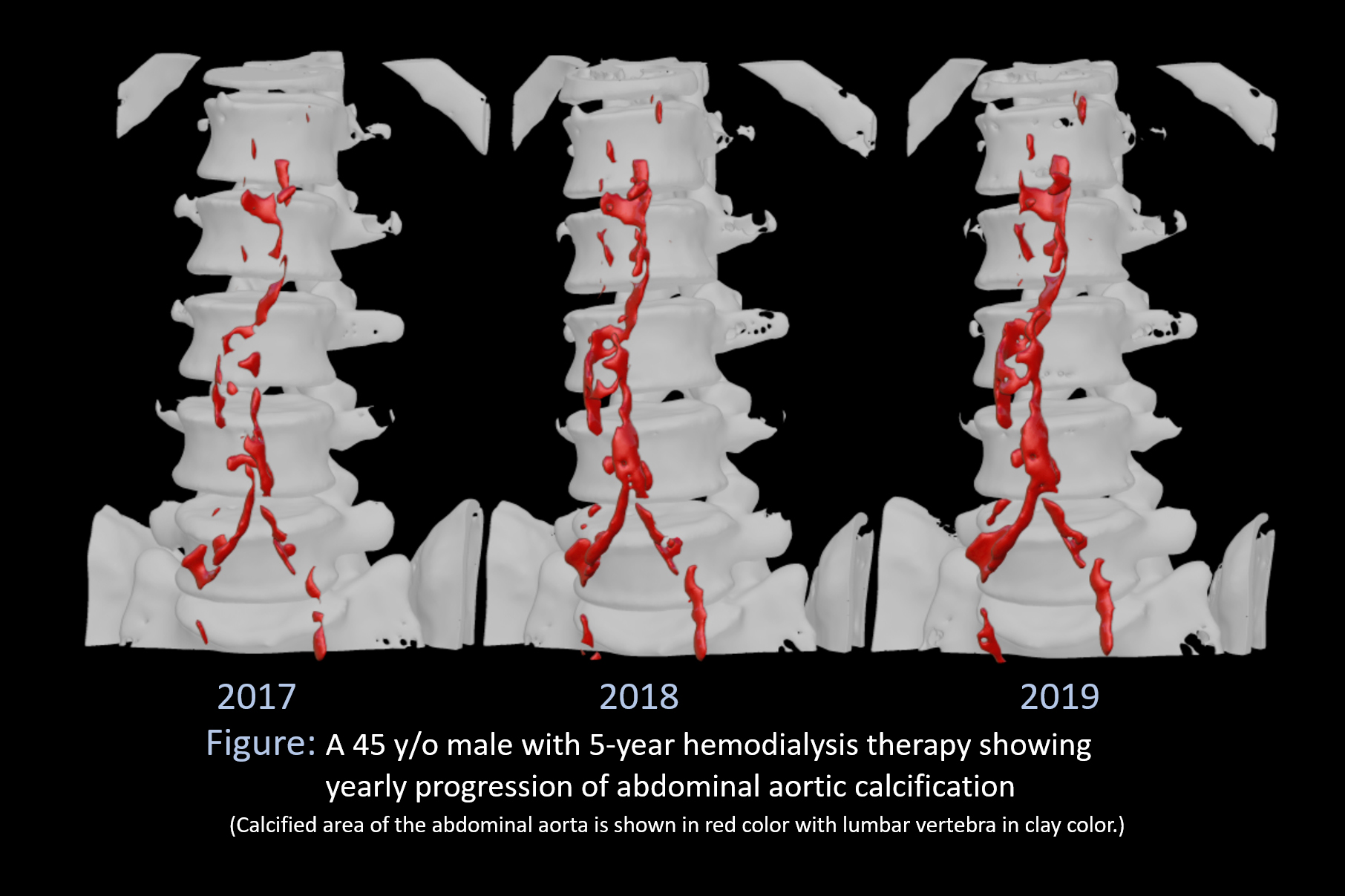
Back to 2020 ePosters