
Ensuring Complete Filter Retrieval Using Ex-vivo Cone-beam Ct Imaging
Kavya Sinha, MD, Ponraj Chinnadurai, PhD, Philip Auyang, MD, Pooja Tekula Reddy, MD, Travis J. Vowels, MD, Alan B. Lumsden, MD.
Houston Methodist, Houston, TX, USA.
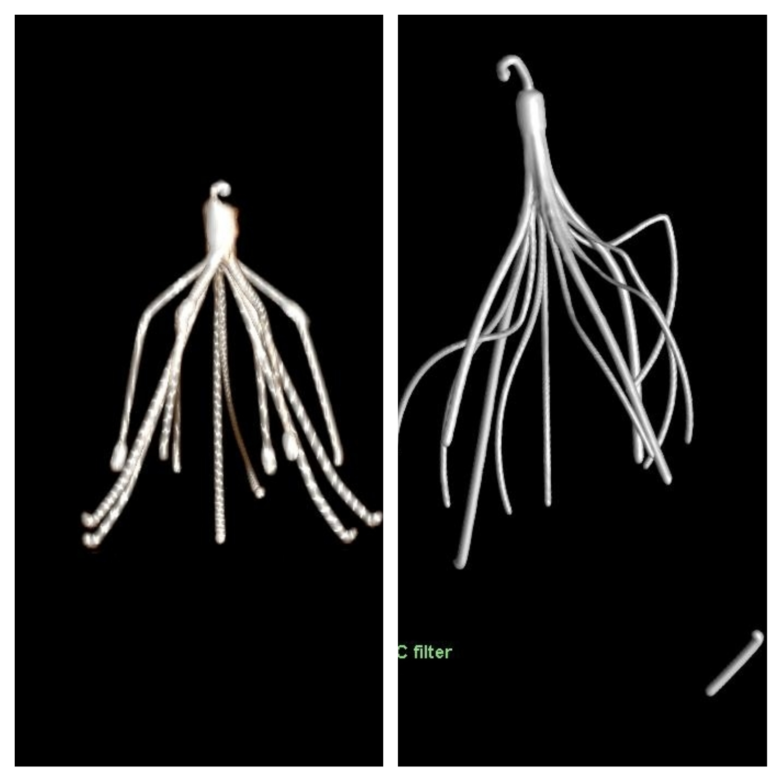
OBJECTIVES: Removal of IVC filters is increasing in frequency due to concerns for long term complications. Although most of these filters are intact many of them are disrupted prior to explant or become damaged during the process of explanation. Retained filter fragments are at risk of embolization or creating persistence of symptoms such as back pain. We believe that it is imperative that ideally all filter fragments are removed during the time of either open or endovascular retrieval. Frequently the filters are distorted or fragmented during the retrieval process. We advocate a combination of intraoperative imaging and ex vivo filter reconstruction and cone beam CT scan as a means of validation of filter explantation. This is the first report of using such techniques to reconstruct filters. METHODS: Careful pre-retrieval assessment of IVC filter position, any significant degree of filter tilting or of hook, and/or strut epithelialization and caval wall penetration by filter components should be considered using dedicated cross-sectional imaging for procedural planning. In complex cases, the risk for retrieval complications should be carefully weighed against the risks of leaving the filter permanently indwelling.
RESULTS: With the introduction of retrievable inferior vena cava filters, the number being placed for protection from pulmonary embolism is steadily increasing. Despite this increased usage, the true incidence of complications associated with inferior vena cava filters is unknown. This article reviews the known complications associated with these filters and suggests recommendations and techniques for inferior vena cava filter removal.
CONCLUSIONS: In this small series, the experience of using DynaCT image fusion guidance together with a steerable endovascular robotic catheter indicates that such image fusion strategies can enhance intraoperative 2D fluoroscopy by bringing preoperative 3D information about vascular stenosis and/or calcification, angulation, and take off from main vessel thereby facilitating ultimate vessel cannulation.
Back to 2020 ePosters