
Automated Vessel Lumen Identification In B-mode Carotid Ultrasound Images Using Convolutional Neural Networks
Lauren Huntress, MD1, Meiyan Xie, BS2, Randy Shafritz, MD1, William Beckerman, MD1, Saum A. Rahimi, MD1, Usman Roshan, PhD3, Justin Ady, MD1.
1Rutgers Robert Wood Johnson Medical School, New Brunswick, NJ, USA, 2New Jersey Institute of Technology, Newark, NJ, USA, 3New Jersey Institute of Technology, New Brunswick, NJ, USA.
OBJECTIVES: Carotid ultrasound is a screening modality used to aid in the prevention of ischemic stroke in high-risk patients. B-mode ultrasound of the carotid artery identifies the vessel wall, lumen, and potential plaque burden within the artery, but requires qualified technicians and physicians to perform and interpret the study. To date, utilization of automated state of the art machine-learning techniques like deep learning in this realm have been limited. Herein, we describe our initial experience with the use of a deep convolutional neural network called U-Net for lumen identification in carotid ultrasounds images.
METHODS: Carotid ultrasound is a screening modality used to aid in the prevention of ischemic stroke in high-risk patients. B-mode ultrasound of the carotid artery identifies the vessel wall, lumen, and potential plaque burden within the artery, but requires qualified technicians and physicians to perform and interpret the study. To date, utilization of automated state of the art machine-learning techniques like deep learning in this realm have been limited. Herein, we describe our initial experience with the use of a deep convolutional neural network called U-Net for lumen identification in carotid ultrasounds images.
RESULTS: Inclusion of all B-mode ultrasound images (internal carotid artery (ICA), external carotid artery, common carotid artery, and carotid bulb) yielded 89.6% overall accuracy in identifying the vessel lumen. Of important consideration, initial accuracy was only 87.6% when sample size was reduced to 20 patients. When images of only the ICA were selected and evaluated independently, the U-Net model's accuracy increased to 95%.
CONCLUSIONS: Deep learning represents a revolutionary, yet underutilized analytical modality in vascular surgery. Our early results demonstrate the feasibility of automated structural identification and independent interpretation of B-mode images by a convolutional U-Net. Our study suggests that accuracy can be improved with increased sample size and categorization of data prior to evaluation. Future studies developing models to accurately distinguish vessel wall, plaque burden, and subsequent degree of stenosis are warranted. 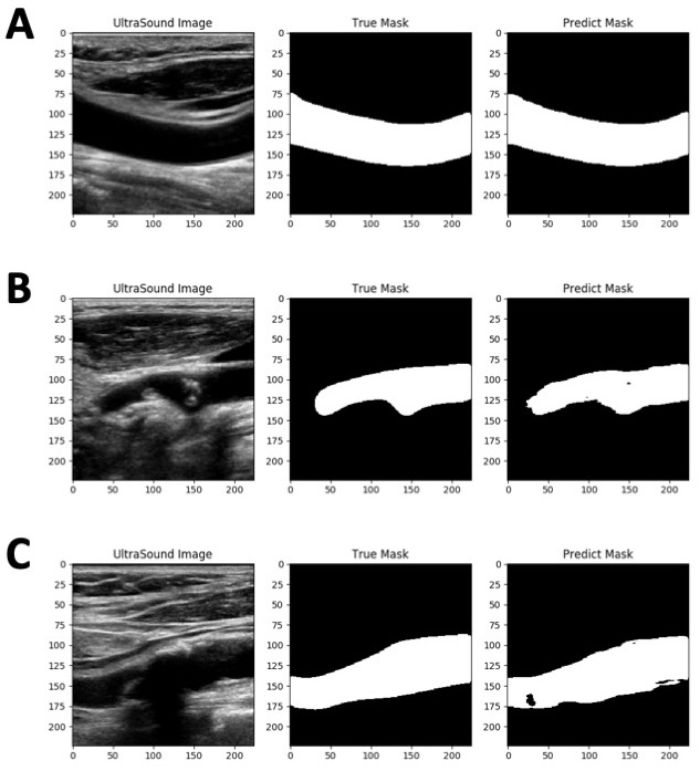
Back to 2020 ePosters