
A Clinically Based Classification System Of The Variations Of The Profunda Femoris Artery And It’S Circumflex Branches.
Alykhan Lalani, MD, Jacob Bray, MD, Claudie Sheahan, MD, Amit Chawla, MD, Malachi Sheahan, MD.
LSU School of Medicine, New Orleans, LA, USA.
OBJECTIVES:The profunda femoris artery (PFA) supplies important collateral branches to both the ipsilateral hypogastric vessel and the distal superficial femoral artery. The size and patency of these collateral pathways can determine the risk of pelvic malperfusion, spinal cord ischemia, and lower extremity limb loss after vascular interventions. Despite its importance, the anatomy of the PFA is rarely characterized in studies involving the hypogastric or lower extremity circulation. This discussion is further limited by the lack of a comprehensive classification system. This study’s aim was to describe the most common PFA anatomic variants and present a classification system based on its branching patterns.
METHODS:In total, 155 cadaveric femoral artery systems were dissected. Notations were made of the configurations of the PFA, lateral circumflex femoral artery (LCFA), and medial circumflex femoral artery (MCFA).
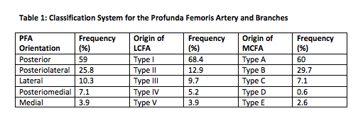
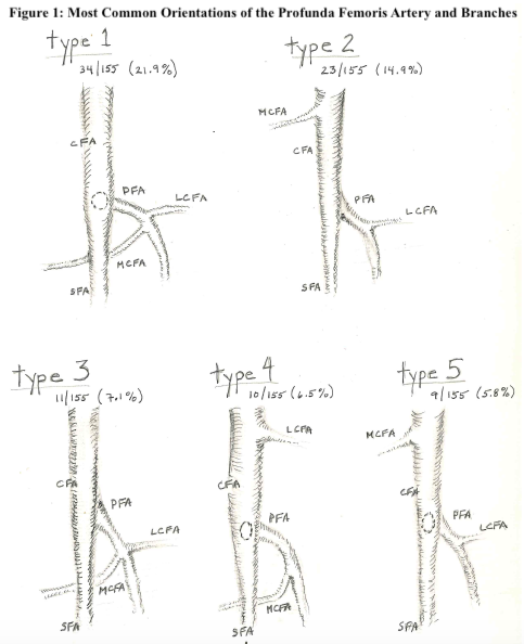
RESULTS:A classification system based on orientation of the PFA, LCFA,and MCFA was created (Table 1). There were five different origins of the LCFA identified. Type I from the PFA, Type II from the CFA, Type III from a shared common origin with the PFA or the MCFA, Type IV from the SFA, and Type V as separate ascending and descending branches from multiple vessels. There were five different origins of the MCFA which were classified as Type A from the PFA, Type B from the CFA, Type C from a common origin between the PFA or the LCFA, Type D from the inferior epigastric or the external iliac above the inguinal ligament, and Type E originating as a double MCFA. While there are 125 potential variants that can be categorized with this system, the five most common types account for 56.1% of the total number seen in our cadaver series (Figure 1).
CONCLUSIONS:The anatomic orientation of the profunda femoris and its branches is highly variable. We propose a classification system that will allow consistency in the terminology used in future studies of open and endovascular interventions which involve the PFA and its collateral beds.
Back to 2021 Abstracts