
Ex-vivo Human Cadaveric Aortic Dissection Model With Flow Circuit For Studying Type A Dissection And Endovascular Repair
Maria Katsarou1, Viony M. Belvroy1, Santi Trimarchi2, Jean Bismuth1.
1Houston Methodist Hospital, Houston, TX, USA, 2University of Milan, Milan, Italy.
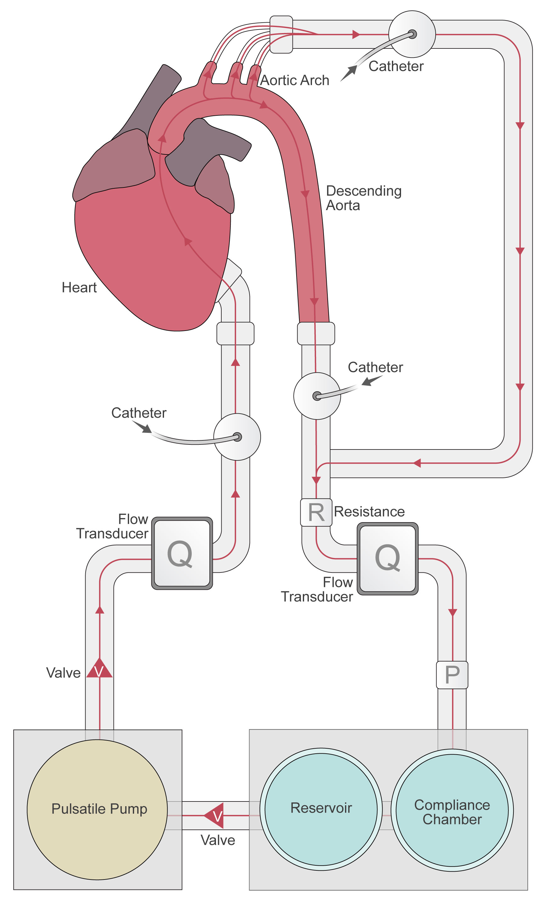
Objectives: Impact of reduced aortic compliance is relevant in the context of endovascular ascending aortic repair and has not been studied extensively. Our objective is to create an ex-vivo human cadaveric aortic dissection model with flow circuit and study the physiological impact of ascending aortic stent grafts using various imaging modalities Methods: Ten cadaveric heart and aortic specimen will be harvested for the experiment. After ligation of all four pulmonary veins, the left ventricle will be perfused using an inflow cannula from pulsatile flow loop construct (HeartBeat Simulator), maintaining physiological aortic valvular flow. Outflow cannula from supra-aortic trunks, and diaphragmatic aorta were connected back to the pump through a compliance chamber and reservoir. The system will be filled with 3 L of blood analogous fluid and run at 60 beats/min to achieve 100-120 mmHg of pressure. An entry tear will be created transapically using an endovascular needle and the dissection flap allowed to progress distally by increasing circuit pressure. Each model will be studied using the following imaging modalities: 2-dimensional phase-contrast MRI, 4D flow MRI, ECG-gated and dynamic CTA, ultrasound (trans-thoracic, intra-cardiac echocardiography). A conformable Gore TAG stent-graft (cTAG, WL Gore and associates, Arizona, USA) will be deployed in the ascending aorta and imaging will be repeated for each model. Results: Our benchtop ex-vivo aortic dissection model, will serve as one of the deductive experimental approaches from swine to human aortas and will enable validation of imaging results from our prior experience with in-vivo swine type B aortic dissection model (figure 2). In our previous experiment, we were able to study spatial and temporal changes using different imaging modalities and we aim to apply the same protocol on ex-vivo aortas under more controllable experimental conditions.Conclusion: This will be, to our knowledge, the first ex-vivo human cadaveric aortic dissection model with flow circuit to study and gain insights into the pathophysiology of type A aortic dissection.However, our ex-vivo model can mimic physiologic hemodynamic conditions like pressure changes and pulsatile flow. Moreover, aortic dimensions, tissue characteristics and histopathology will be closer to real-life human conditions than healthy animal aortas.
Back to 2022 ePosters