
Ultrasound Elastography To Quantify Pressure Normalized Strain Reduction Associated With Different Aortic Endografts In A 3D-printed Hydrogel Phantom
Dakota W. Gonring, Bachelor of Arts, Zachary R. Zottola, BS, Maxwell L. Wang, BA, Michael S. Richards, PhD, Michael C. Stoner, MD, Doran S. Mix, MD.
University of Rochester Medical Center, Rochester, NY, USA.
OBJECTIVES: Recent literature has shown that strain may be useful in evaluating stability of abdominal aortic aneurysms (AAA) following endovascular aneurysm repair (EVAR). With the numerous options available for choice of endograft and little data to assess the efficacy of different graft compositions, it is useful to quantify the magnitude of strain reduction associated with different grafts. Here, we measure pressure normalized strain associated with different endografts in a hydrogel phantom using our novel ultrasound elastography (USE) technique.
METHODS: A homogenous 10% by mass polyvinyl alcohol cryogel (PVA-c) 3D-printed phantom (Figure 1.A) with a 28 mm diameter was connected to a cardiac flow simulator and imaged after implantation with four different 30 mm EVAR grafts using USE (Figure1.B). Graft compositions were nitinol/platinum-iridium/polyester (A), polyester/stainless steel (B), PTFE/nitinol (C), and polyester/electropolished nitinol (D). Mean principal strain (ep+) was measured three times in the main body of each graft. The strain values for each trial were then normalized by dividing by pulse pressure to yield pressure-normalized strain (ep+/pp). This procedure was repeated for the phantom with no endograft in place (NOG). Separate paired two tailed T-tests were performed comparing ep+/pp in the phantom with no graft to the different ep+/pp values obtained from the phantom with each individual graft.
RESULTS: There was a statistically significant reduction in the average ep+/pp for each graft compared to the phantom with no graft (NOG: 0.946 ± 0.019%/kPa, D: 0.739 ± 0.047%/kPa; p= 0.028, B: 0.733±0.067%/kPa; p= 0.040, C: 0.649±0.086%/kPa; p= 0.035, A: 0.599±0.031%/kPa; p= 0.006). There was a significant difference between graft D and graft A (p=0.013) ep+/pp which was not present for the other grafts.
CONCLUSIONS: This study demonstrates the ability of USE to measure ep+/pp associated with different endografts in a 3D-printed vessel phantom. Our data suggest that there is a significant difference in the absolute value of strain between graft manufacturers. Further exploration is needed to determine if this is clinically significant. 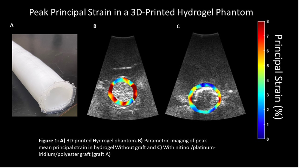
Back to 2022 ePosters