
MRI Characterization Of Popliteal And Tibial Plaques In Diabetic Patients To Aid Patient Selection For Limb Salvage Endovascular Treatments
Kavya Sinha, MD1, Christof Karmonik, PhD2, Alan Lumsden, MD1, Trisha Roy, MD, PhD1.
1Houston Methodist, Houston, TX, USA, 2Houston Methodist Academic Institute, Houston, TX, USA.
OBJECTIVES:Diabetic patients with limb ischemia are at high risk of major amputation. The “endovascular-first” strategy has been widely adopted for limb salvage, but not all lesions are amenable to endovascular treatment. Our objective is to use MRI to characterize popliteal and tibial plaques in complex diabetic patients to identify plaques that are at high risk of endovascular failure.
METHODS:8 diabetic patients undergoing endovascular limb salvage procedures were studied. Patients were imaged on a 3T MRI scanner at 0.5 mmł isotropic resolution and scanned with an Ultrashort Echo Time (UTE) sequence to identify hard components (calcium and collagen), and a Steady State Free Precession (SSFP) sequence to identify soft components, as previously described. MR images were correlated with pre-operative duplex ultrasound +/- CTA, and intraprocedural X-ray angiography images.
RESULTS:Endovascular treatments were performed by three vascular surgeons at a large academic referral hospital. 6/8 patients had prior endovascular treatment from an outside hospital and 5/8 had end-stage renal disease (ESRD). All 3 surgeons aborted at least 1 endovascular case. 4/8 (50%) of procedures were aborted in total, 3 because of inability to cross a hard lesion, and 1 due to discrepancy between ultrasound and angiogram regarding complexity of the lesion. MR images demonstrated soft lesion components in all 4/4 patients that had successful interventions. MR images demonstrated dense collagen and calcium in all 4/4 patients that failed intervention. Of note, the dense collagen was not seen on CTA or X-ray angiogram and there was minimal calcification in 3 / 4 patients who failed intervention (Figure 2).
CONCLUSIONS:
Diabetic patients with ESRD and prior interventions have a very high risk of endovascular failure. Many patients would benefit from bypass surgery or amputation, but without clarity on patient selection, many endovascular treatments are still attempted. MRI can identify dense collagen lesions that are at high risk of endovascular failure and these high-risk lesions are not seen on current clinical imaging modalities. This work demonstrated how MRI plaque characterization offers important information to aid patient selection for endovascular limb salvage treatments in the future. 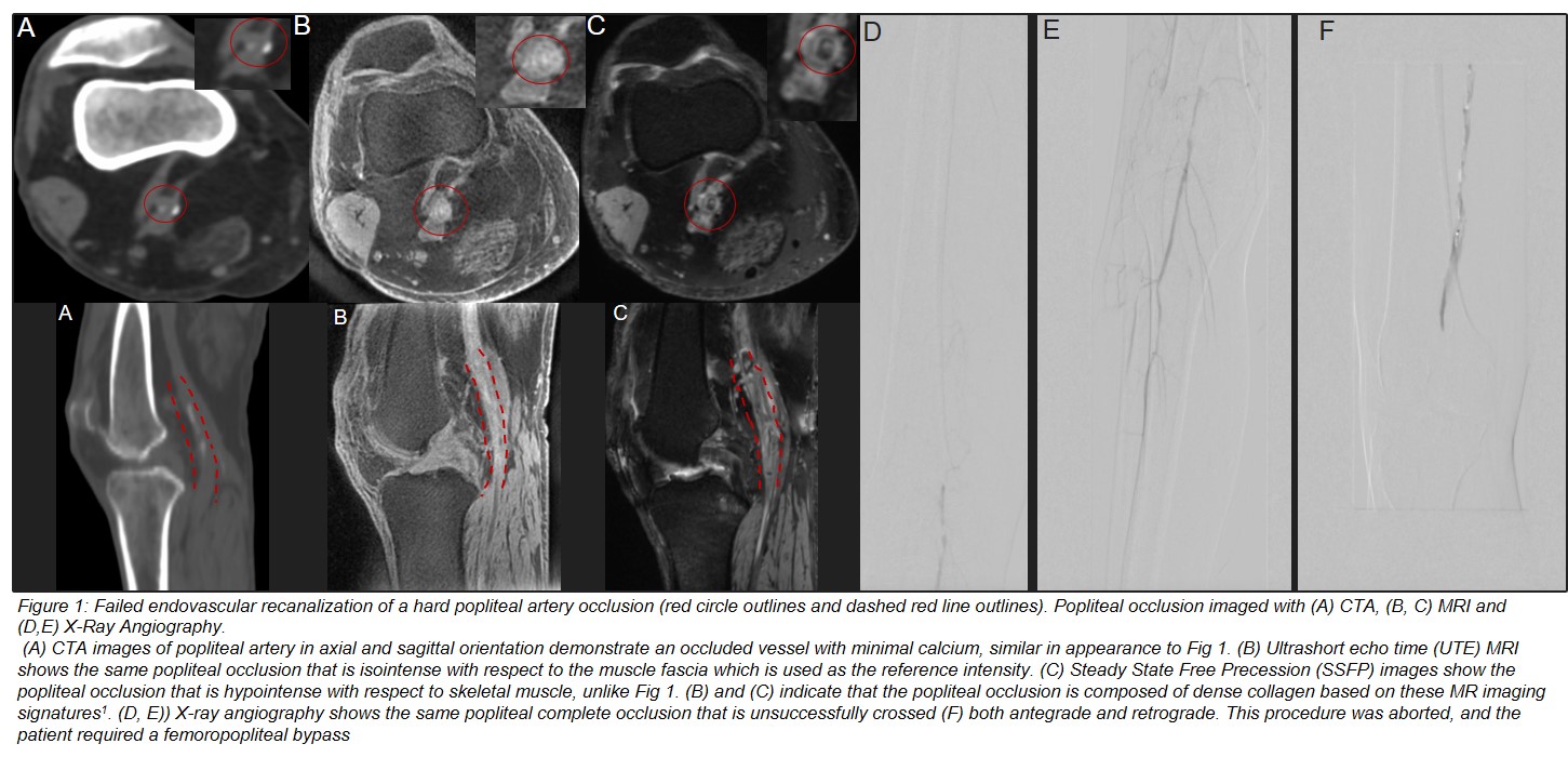
Back to 2022 ePosters