
Fabric Tears And Type IIIb Endoleaks After Fenestrated-branched Endovascular Aortic Aneurys Repair (F-BEVAR)
Jesus Porras Colon, MD, Carla K. Scott, MD, Felipe Pavarino, MD, Marilisa Soto Gonzalez, MD, Khalil H. Chamseddin, MD, Mirza S. Baig, MD, Melissa L. Kirkwood, MD, Carlos H. Timaran, MD.
UT Southwestern, Dallas, TX, USA.
Background: Type IIIb endoleaks from fabric tears after F-BEVAR are rare. The aim of this study was to describe the incidence and management of Type IIIb endoleaks F-BEVAR.
Methods: A total of 268 patients undergoing F-BEVAR with investigational devices between 2015 and 2021 were analyzed. All patients with type IIIb endoleaks were included. End points were presentation of type IIIb endoleaks and secondary interventions.
Results: Three patients developed type IIIb endoleaks (2 females, mean age 75 years). The first patient was a 74-year-old female with underwent F-BEVAR for a thoracoabdominal aortic aneurysm with a device with two branches and two fenestrations. The 4-year postoperative CT angiography (CTA) demonstrated an endoleak behind the branches, which was confirmed to be a fabric tear with dynamic volumetric CTA (DV-CTA)(Fig). The patient underwent a successful redo-F-BEVAR. The second patient was a 74-year-old female that underwent branched repair with a device with four branches and an inverted limb bifurcated device after a failed endovascular repair. On the 30-day follow-up CTA, an endoleak of indeterminate origin was detected. Further imaging, including CTA, DV-CTA and diagnostic angiogram confirmed a tear in the graft posterior to a renal branch. A redo branched repair was successfully performed. The last case was a 78-year-old male who underwent a 4 vessel F-BEVAR with an inverted limb custom-made device after failed endovascular repair. On the 30-day follow up visit, a type IIIb endoleak around the crutch of the inverted limb device was identified by CTA, contrast enhanced ultrasound and color-Doppler ultrasound. The defect was excluded with an iliac limb extension. Technical success of the reintervention was a 100% with no deaths. Sac growth prior to reintervention was identified in all cases. Mean follow-up was 30 months. The endoleak development occurred on an average of 17 months after index operation. The overall frequency of type IIIb endoleaks in our series was 1.1 %.
Conclusion: Type IIIb endoleaks after F-BEVAR are rare. Management of these cases is technically challenging. Proper multimodality imaging and redo F-BEVAR are crucial for proper diagnosis and treatment of this rare complication. 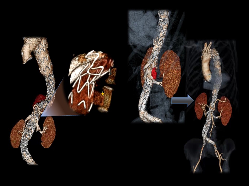
Back to 2022 ePosters