Back to 2024 Display Posters
Shaggy Aorta Assessment With Intravascular Ultrasound Prior To Fenestrated And Branched Endovascular Aortic Aneurysm Repair
Mira T. Tanenbaum, MD, Marilisa Soto Gonzalez, MD, Andres V. Figueroa, MD, Jose Eduardo Costa Filho, MD, Mirza S. Baig, MD, Carlos H. Timaran, MD.
University of Texas Southwestern Medical Center, Dallas, TX, USA.
OBJECTIVES:Fenestrated and branched endovascular aortic aneurysm repair (FBEVAR) has emerged as an effective treatment option for complex aortic pathologies. Patients with shaggy aortas, characterized by severe aortic atherosclerosis with irregular and ulcerated plaques, pose unique challenges given the increased risk of perioperative embolic complications. Ultrasound may be used to assess atherosclerotic plaque composition to predict adverse embolic events. The utility of intravascular ultrasound (IVUS) in patients with shaggy aorta undergoing FBEVAR was assessed.
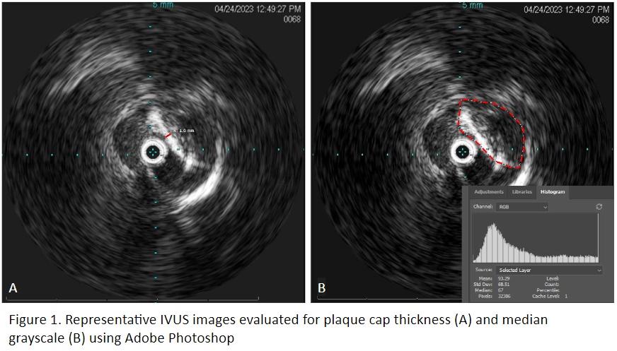
METHODS: Consecutive FBEVAR procedures in patients with shaggy aortas between 2018 and 2023 as part of a physician-sponsored investigational device exemption study were reviewed. IVUS was performed to assess echogenicity and stability of the aortic plaque prior to considering repair. Perioperative data, including technical success, rates of embolization events, mortality, major complications, and long-term outcomes were assessed. IVUS images were used to determine echogenicity, cap thickness and median grayscale values.
RESULTS: Among 1330 patients undergoing FBEVAR, 5 (0.38%) patients with shaggy aortas underwent FBEVAR at a single center. All patients were female (mean age of 73 years) with a smoking history, hypertension, and hyperlipidemia. The median maximal aneurysm diameter was 63 mm (53-63). All 5 patients were on preoperative statin therapy; 3 (60%) patients were taking aspirin. A total of 17 fenestrations were planned. Staged TEVAR was performed in 3 (60%) patients prior to FBEVAR. A distal atheroembolic event during the staged TEVAR procedure necessitating hallux amputation occurred in 1 (33%) patient. Technical success was 100% with no instances of embolic events, visceral or lower extremity malperfusion, or vascular injury after FBEVAR. IVUS imaging analysis showed median cap thickness of 0.8 mm (0.6-1). Plaques generally appeared stable and echogenic with median grayscale value of 104 (69.5-112.5) (figure). Primary patency was 100% at median follow-up time of 42.5 days (31.5-351.75). During follow-up, three (60%) type II endoleaks were detected with no secondary interventions indicated.
CONCLUSIONS: FBEVAR in patients with shaggy aortas is feasible. IVUS may aid in determining the safety of FBEVAR by evaluating plaque cap thickness and echogenicity. Further studies are needed to determine the benefits of IVUS plaque evaluation in shaggy aortas.

Back to 2024 Display Posters