Back to 2024 Display Posters
The Use Of Pulse Wave Doppler To Characterize Embolic Events In A Flow Loop System
Basil P. Alias, B.S.1, Divek U. Toprani, MTM
1, Emily Lo, B.S.
1, Zsolt Garami, MD
2, Stuart J. Corr, PhD
2, Alan B. Lumsden, MD
2.
1Texas A&M School of Engineering Medicine (ENMED), Houston, TX, USA,
2Houston Methodist Hospital, Houston, TX, USA.
OBJECTIVES: Carotid embolectomies and endarterectomies (CEA) previously required large invasive surgeries to prevent perioperative embolization from working sites. Modern approaches including flow reversal and filters still fail to prevent post-CEA strokes, raising questions regarding the effectiveness of filters and whether post-CEA infarcts might form via alternate mechanisms. The purpose of this study is to further explore the capabilities and limitations of emboli detection using pulse wave doppler ultrasound (US) with the ultimate goal of continuous noninvasive characterization of embolic events intra-operatively.
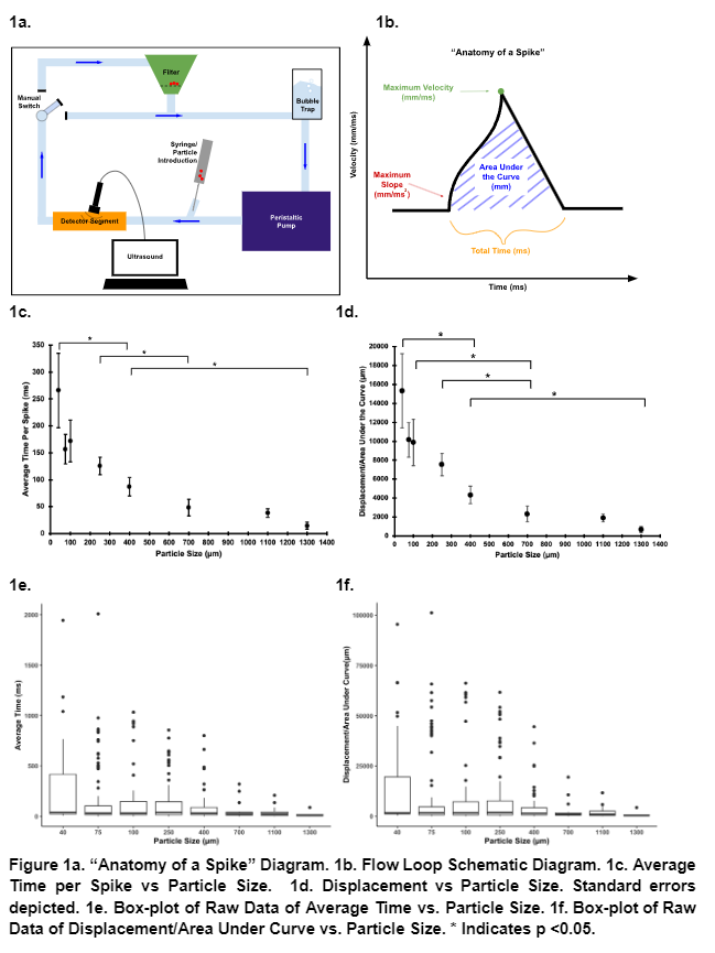
METHODS: An experimental flow loop system was devised to mimic embolus movement within blood vessels. This setup ensures emboli integrity while enabling solution circulation, contaminant removal, controlled emboli introduction, and emboli detection via ultrasound (Spencer Technologies PMD100). Baseline flow data is collected for 30 seconds at 200 RPM (226 mL/min) using the Kamoer KCM pump. Subsequently, 0.2 mL of Embozene Microspheres is injected, circulating for 1 minute for particle distribution and bubble removal. Another 30-second flow dataset is collected before a 1-minute filtration in preparation for subsequent tests. This cycle is repeated thrice for emboli of sizes: 40 μm, 75 μm, 100 μm, 250 μm, 400 μm, 700 μm, 1100 μm, and 1300 μm. Analysis employs MATLAB R2023a, R statistical software, and Microsoft Excel.
RESULTS: For each test 30 seconds of baseline and emboli data were collected. Filtering out baseline flow noise from the emboli data revealed velocity spikes produced by emboli passing the US transducer. These spikes were then individually identified and analyzed. For each spike 4 parameters were collected: Maximum Slope, Maximum Velocity, Area Under the Curve, and Total Time (Figure 1). Parameters were compared with a t-test between sample averages. Results of the study indicated that particle size varies inversely with both Displacement and Average Time per Spike.
CONCLUSIONS: The study results suggest that pulse-wave doppler has potential to be used to characterize microemboli down to 40 µm. Next steps include testing carotid filters on the system and correlating data in vivo based on the regression equations.

Back to 2024 Display Posters