DEMOGRAPHICS:69-year-old White female. HISTORY:Presented with multidrug resistant hypertension and worsening kidney disease. She has a history of coronary and peripheral artery disease status post aortobiiliac bypass in 1994 that was complicated by occlusion of the left aortoiliac limb, requiring right to left femoro-femoral crossover graft in 2007. She had redo crossover bypass in 2009 and right femoral endarterectomy in 2017. Computed tomography angiogram showed diffuse circumferential calcification of the aortic arch to the aortic bifurcation, coral reef plaque causing occlusion of the left renal artery (LRA) with atrophic left kidney, stenosis of right renal artery (RRA) and superior mesenteric (SMA), and a patent right aortoiliac limb and right to left femoro-femoral graft. Her ankle-brachial indices were 0.57 bilaterally, and echocardiogram showed an ejection fraction of 70% with normal valvular function.
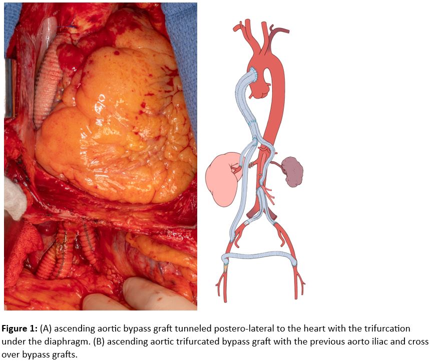
PLAN:Via a redo laparotomy, exposure of the supraceliac aorta, RRA, SMA and right external iliac artery (REIA) was achieved. The ascending aorta was exposed via a median sternotomy, a side-biting clamp was placed on its anterolateral surface and an end-to-side anastomosis was done using a 14mm woven Dacron graft. This was tunneled through an opening in the diaphragm coursing medial to the mobilized left lobe of the liver. A trifurcated graft was fashioned on the back table by sewing a 7mm Dacron limb end-to-side to one of the 8mm limbs of a 16x8mm bifurcated Dacron graft. The trifurcated graft was sewn in an end-to-end fashion with the ascending aortic graft. Retroperitoneally, one of the 8mm limbs was sewn end-to-side to the REIA and the 7mm limb was sewn end-to-end to the RRA. The other 8mm limb was tunneled in a retropancreatic fashion and sewn end-to-side with the SMA. Intraoperative Doppler signals were found in the RRA, SMA, and feet. She progressed well postoperatively despite developing acalculous cholecystitis that was treated with percutaneous cholecystostomy tube placement. Patient was discharged home on post-operative day 16 and is doing well after 2 months with controlled blood pressure and improved kidney function.
DISCUSSION:The ascending aorta can be used as an inflow source in the setting of diffuse aortic calcification precluding more distal aortic control.