A Morphometric Analysis of the Impact the Cardiac Cycle has on Thoracic Aortic Geometry
Muhammad M. Zubair, MD, Adeline Schwein, MD, Ali Irshad, MD, Carlos F. Bechara, MD, Faisal Nabi, MD, Jean Bismuth, MD.
Houston Methodist Hospital, Houston, TX, USA.
Objective: Endovascular thoracic aortic disease management, particularly in the ascending and aortic arch, are likely to be greatly impacted by morphological changes during the cardiac cycle. The aim of our study was to assess these geometrical variations.
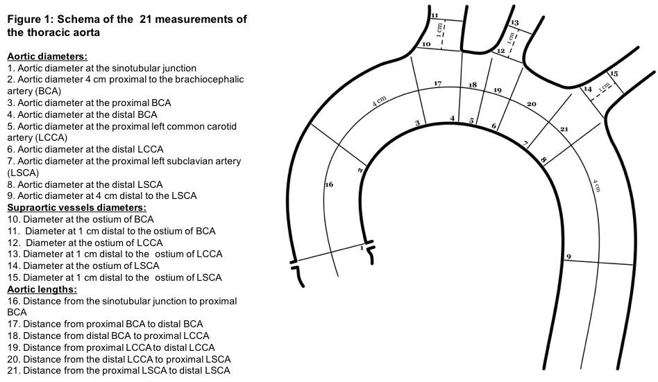
Methods: In this retrospective study, we included 8 patients who underwent an EKG-gated Multi-Detector Computed Tomography (MDCT). Subjects with ejection fraction (EF) ≥ 45% were selected. We defined both end-systolic and end-diastolic MDCT phases by assessing the lowest and highest left ventricular volumes respectively. We established 21 measurements of maximal diameters and lengths of the ascending, arch and descending aorta as well as the origin of the supra-aortic vessels (Figure 1). Measurements were made perpendicular to the centerline on both systolic and diastolic phases, independently by two physicians, using the software Syngo.Via (Siemens Healthineers). Inter-rater reliability was evaluated using the intraclass correlation coefficient (ICC), while a paired t-test was used to assess differences between systole and diastole within a single rater’s measurements.
Results: Our 8-patient cohort included 6 males with a mean (±SD) age of 70.12 years old (± 11.4) and a mean EF of 62.15% (± 9.9). The ICC evaluation showed good correlation (lower limit of the 95% confidence interval >0.75) for all diameter measurements involving the aorta (#1 to #9 in figure 1). Higher variability was found for aortic lengths and measurements of the supra-aortic vessels. Regarding aortic diameters, they were all larger in the systolic phase when compared to the diastolic phase, with significant differences for 5 of them (5/10). Aortic lengths of the ascending aorta and the proximal arch were also significantly longer in the systolic phase.
Conclusion: Our results show significant variations of diameters and lengths of the thoracic aorta along the cardiac cycle. These findings allow for a better understanding of morphometric variation during the cardiac cycle, which should especially be taken into consideration for future designs and sizing of endovascular devices.
Back to 2017 Karmody Posters


